262021 A choroid plexus cyst is a cyst that can grow in the brain of a fetus during development. Today I got a call from my regular OB who said she needed to schedule me for a fetal echocardiogram and.
 Chromosomal Anomalies Radiology Key
However the risk when no associated anomalies are detected is much less well defined.
Choroid plexus cyst in fetus. An abnormal karyotype was identified in four 64 of these fetuses. However in a very small percentage of fetuses with choroid plexus cysts there is an associated chromosome disorder called trisomy 18. 412013 A choroid plexus cyst is a small fluid-filled structure within the choroid of the lateral ventricles of the fetal brain.
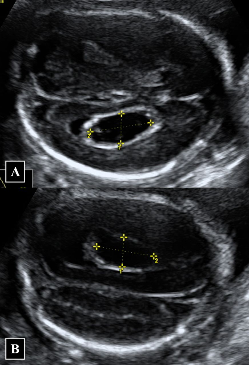
Ive lost 3 1 was ectopic my ds 20mths old was born with ectrodactyly and 7 weeks premature. However in small number of fetuses choroid plexus cyst may be a marker for genetic disorder called trisomy 18. To identify a chroid plexus cyst CPC it must be imaged in two orthogonal planes and be greater than or equal to 3 mm in size.
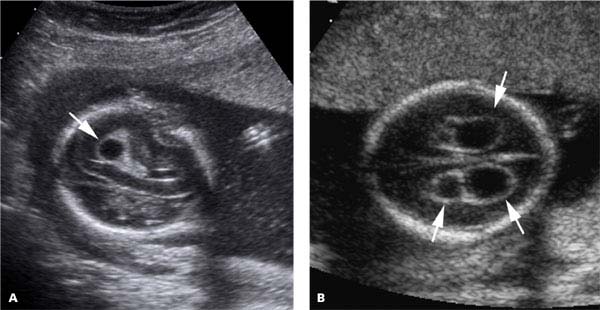
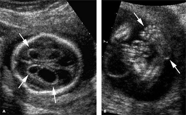
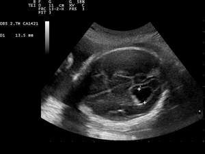
A choroid plexus cyst is a small fluid-filled structure within the choroid of the lateral ventricles of the fetal brain. More than 90 resolve by 26 weeks. Single or multiple cystic areas 2 mm in diameter in one or both choroid plexuses of the lateral cerebral ventricles.
3252019 Choroid Plexus Cysts CPC are small fluid filled areas in the brain and they are a common ultrasound finding in the fetus during the 2nd trimester of pregnancy. Im hoping someone out there has had experience with CPCs choroid plexus cyst and can share some experiences. 11262012 Choroid plexus cyst on its own may not have an impact on the health of fetus its growth and development or learning after the birth of the fetus.
They are most commonly seen in the second and third trimester of the pregnancy and can be detected via an ultrasound. 312012 Choroid Plexus Cyst found on 20 week Ultrasound. However when no other anomalies are seen on the ultrasound the risk of a change in the number of chromosomes is still around 1 in 300.
The prevalence of fetal choroid plexus cysts was 08 172084 during a 40-month period. Sixty-two of the 71 patients elected to undergo amniocentesis. All cysts were initially identified on sonograms performed between 14 and 21 weeks.
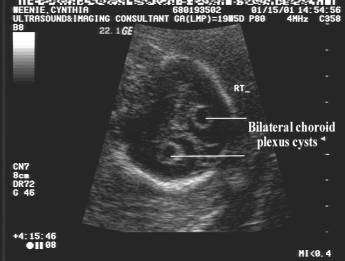
1 in 50 fetuses at 20 weeks gestation. Sonographically choroid plexus cysts appear as echolucent cysts within the echogenic choroid Figure 1. Cysts ranged from 3 to 11 mm in size and were bilateral in four 36 of.
I consulted a second perinatologist and radiologist as well as a pediatric neurologist all of whom informed me that in a baby with normal chromosomes choroid plexus cysts usually disappear in the 2nd-3rd trimester and it is unknown if they are clinically important. A choroid plexus cyst is not considered a structural or functional brain abnormality. When associated anomalies are detected the risk is sufficient to justify an invasive diagnostic test such as amniocentesis.
Fetal choroid plexus cysts were diagnosed in 71 096 of these pregnancies. The doctor told me that the baby had a choroid plexus cyst in her brain and told me it was nothing to worry about because I was cleared in my NIPT testing for genetic conditions and that choroid plexus cysts are normally not an issue. Fetuses with trisomy 18 have an extra copy of chromosome 18.
One fetus had trisomy 21 and three fetuses were diagnosed with trisomy 18. This 5th pregnancy was going great until. In fetuses with choroid plexus cysts 21 have an abnormal number of chromosomes with the majority having other anomalies that can be seen on the ultrasound.
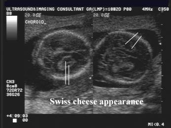
Choroid plexus CP cysts are commonly detected on routine mid-trimester ultrasound scan. Choroid plexus cysts may be single or multiple unilateral or bilateral and most often are less than 1 cm in diameter. Sonographically choroid plexus cysts appear as echolucent cysts within the echogenic choroid Figure 1.
Screening ultrasonographic examinations were performed on 16059 patients and 301 patients had a fetus with a choroid plexus cyst. A 20-year-old woman at 18 weeks period of gestation was referred to the fetal medicine clinic in view of ultrasound USG finding of large irregular bilateral choroid plexus cysts CPCs measuring 16279 mm in the right lateral ventricle and 14474 mm in the left lateral ventricle. In most instances these are a normal variant.
Frequently fetuses with trisomy 18 are stillborn. This is my 5th pregnancy. After a screening ultrasonography.
One hundred thirty patients elected to have an. As mentioned choroid plexus cysts are present in 1 to 2 percent of normal fetuses. Any patient with a choroid plexus cyst was offered genetic counseling and an amniocentesis.
 Prenatal Ultrasound Findings A Choroid Plexus Cysts At 18 Weeks Of Download Scientific Diagram
 Choroid Plexus Anomalies Cysts And Papillomas Sciencedirect
 Isolated Large Bilateral Choroid Plexus Cysts Associated With Trisomy 18 Bmj Case Reports
 Isolated Fetal Choroid Plexus Cysts
 Choroid Plexus Cyst Wikipedia
Https Mydoctor Kaiserpermanente Org Ncal Images Gen Us 20cpc 20handout Tcm63 15061 Pdf
 Medpix Case Trisomy 18
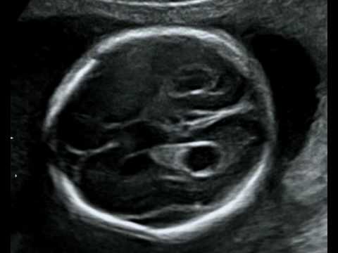 Fetal Medicine Foundation Choroid Plexus Cyst Youtube
 Choroid Plexus Cyst Radiology Case Radiopaedia Org
Chromosomal Anomalies Radiology Key
 Sagittal Plane Of The Fetal Head Shows A Choroid Plexus Cyst Download Scientific Diagram
 Choroid Plexus Cyst Cpc Hkog Info
Comments
Post a Comment